Copyright
©The Author(s) 2015.
World J Radiol. Jun 28, 2015; 7(6): 110-127
Published online Jun 28, 2015. doi: 10.4329/wjr.v7.i6.110
Published online Jun 28, 2015. doi: 10.4329/wjr.v7.i6.110
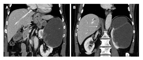
Figure 13 The 70-year-old man with advanced-stage papillary renal cell carcinoma of the left kidney.
Coronal multiplanar reformations during the nephrographic phase show large, mainly cystic left renal mass, with solid contrast-enhancing components (arrowheads). Enlarged retroperitoneal LNs, inhomogeneously enhancing (arrow, A) are detected, compatible with metastatic lymphadenopathy. Liver (arrowhead, B) and right adrenal (long arrow, B) metastases are also seen. All metastatic deposits have a similar pattern of enhancement.
- Citation: Tsili AC, Argyropoulou MI. Advances of multidetector computed tomography in the characterization and staging of renal cell carcinoma. World J Radiol 2015; 7(6): 110-127
- URL: https://www.wjgnet.com/1949-8470/full/v7/i6/110.htm
- DOI: https://dx.doi.org/10.4329/wjr.v7.i6.110


