Copyright
©The Author(s) 2015.
World J Radiol. Jun 28, 2015; 7(6): 110-127
Published online Jun 28, 2015. doi: 10.4329/wjr.v7.i6.110
Published online Jun 28, 2015. doi: 10.4329/wjr.v7.i6.110
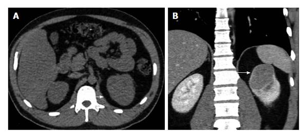
Figure 9 The 31-year-old man with chromophobe renal cell carcinoma of the left kidney (stage T1b, grade II).
A: Axial plain image barely depicts upper pole left renal mass mainly isodense, with a slight bulging of the renal contour (arrow); B: Coronal reformation during the nephrographic phase clearly depicts left renal tumor (arrow). The neoplasm enhances moderately and homogeneously [computed tomography (CT) density: 70 HU, when compared to the CT density of 35 HU on unenhanced images]. A thin hyperdense rim surrounds renal malignancy, proved to correspond to fibrous pseudocapsule histologically.
- Citation: Tsili AC, Argyropoulou MI. Advances of multidetector computed tomography in the characterization and staging of renal cell carcinoma. World J Radiol 2015; 7(6): 110-127
- URL: https://www.wjgnet.com/1949-8470/full/v7/i6/110.htm
- DOI: https://dx.doi.org/10.4329/wjr.v7.i6.110


