Copyright
©The Author(s) 2015.
World J Radiol. Jun 28, 2015; 7(6): 110-127
Published online Jun 28, 2015. doi: 10.4329/wjr.v7.i6.110
Published online Jun 28, 2015. doi: 10.4329/wjr.v7.i6.110
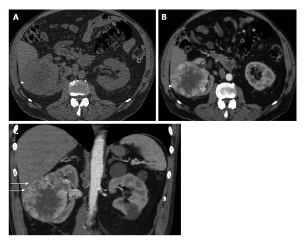
Figure 8 The 75-year-old man with clear cell renal cell carcinoma of the right kidney, invading the liver.
A: Axial plain image shows right heterogeneous right renal tumor (arrowhead); B: Transverse reformation during the corticomedullary phase depicts strong, heterogeneous mass enhancement. The tumor (arrowhead) enhances mainly in the periphery, with a mean computed tomography density of 110 HU (compared to that of 40 HU on the unenhanced images), a finding more compatible with the diagnosis of renal cell carcinoma of the clear cell variety. Central non-enhancing areas corresponded to areas of necrosis on pathology; C: Coronal reformation during the same phase shows renal tumor invading the liver (small arrows), a finding confirmed both on surgery and histopathology.
- Citation: Tsili AC, Argyropoulou MI. Advances of multidetector computed tomography in the characterization and staging of renal cell carcinoma. World J Radiol 2015; 7(6): 110-127
- URL: https://www.wjgnet.com/1949-8470/full/v7/i6/110.htm
- DOI: https://dx.doi.org/10.4329/wjr.v7.i6.110


