Copyright
©The Author(s) 2015.
World J Radiol. Jun 28, 2015; 7(6): 110-127
Published online Jun 28, 2015. doi: 10.4329/wjr.v7.i6.110
Published online Jun 28, 2015. doi: 10.4329/wjr.v7.i6.110
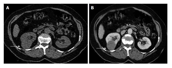
Figure 7 The 60-year-old woman with clear cell renal cell carcinoma of the left kidney (stage T1a, grade II).
A: Transverse plain computed tomography (CT) image depicts lower pole right renal mass as a focal bulging of the renal contour (arrow), mainly isodense (CT density: 35 HU) to the renal parenchyma; B: Axial multiplanar reformation during the corticomedullary phase clearly shows renal malignancy (arrow) moderately and heterogeneously enhancing (mean CT density: 60 HU). Heterogeneous contrast enhancement on imaging should always suggest renal malignancy preoperatively.
- Citation: Tsili AC, Argyropoulou MI. Advances of multidetector computed tomography in the characterization and staging of renal cell carcinoma. World J Radiol 2015; 7(6): 110-127
- URL: https://www.wjgnet.com/1949-8470/full/v7/i6/110.htm
- DOI: https://dx.doi.org/10.4329/wjr.v7.i6.110


