Copyright
©The Author(s) 2015.
World J Radiol. Jun 28, 2015; 7(6): 110-127
Published online Jun 28, 2015. doi: 10.4329/wjr.v7.i6.110
Published online Jun 28, 2015. doi: 10.4329/wjr.v7.i6.110
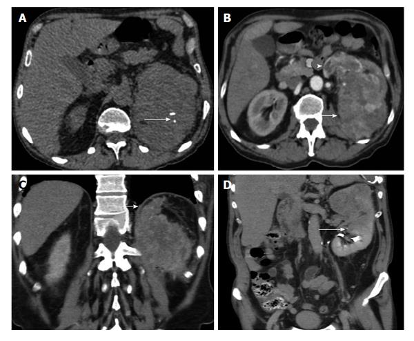
Figure 5 The 62-year-old man with clear cell renal cell carcinoma of the left kidney (stage T3a, grade 3).
A: Transverse unenhanced computed tomography (CT) image shows large heterogenous left renal mass, with small areas of calcifications (long arrow); B: Transverse multiplanar reformation (MPR) during the corticomedullary phase demonstrates left renal malignancy (arrow), inhomogeneously enhancing. The left renal vein is dilated and enhances heterogeneously (arrowhead) due to neoplastic invasion. VTT enhances with a same pattern as renal cell carcinoma; C: Coronal reformations during the corticomedullary phase depicts tumor ill-defined margins and extension into the perinephric fat tissue (arrow). Thickening of the diaphragms of the perinephric space is also seen; D: Coronal MPR during the excretory phase shows nonvisualization of the upper calyces and invasion of the middle calyceal group (arrow), a finding strongly suggestive of invasion of renal sinus fat. CT findings were confirmed both surgically and pathologically.
- Citation: Tsili AC, Argyropoulou MI. Advances of multidetector computed tomography in the characterization and staging of renal cell carcinoma. World J Radiol 2015; 7(6): 110-127
- URL: https://www.wjgnet.com/1949-8470/full/v7/i6/110.htm
- DOI: https://dx.doi.org/10.4329/wjr.v7.i6.110


