Copyright
©The Author(s) 2015.
World J Radiol. Jun 28, 2015; 7(6): 110-127
Published online Jun 28, 2015. doi: 10.4329/wjr.v7.i6.110
Published online Jun 28, 2015. doi: 10.4329/wjr.v7.i6.110
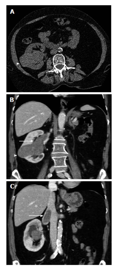
Figure 1 The 65-year-old woman with papillary renal cell carcinoma of the right kidney and tumoral invasion of the ipsilateral renal vein and the inferior vena cava (stage T3b, grade 2).
The patient had left radical nephrectomy years ago for renal cell carcinoma. A: Transverse unenhanced computed tomography (CT) image shows a lobular right renal mass (arrowhead), located in the interlobar region. The mass is relatively homogeneous, slightly hyperdense (CT density: 40 HU), when compared to the normal renal parenchyma; B and C: Contrast-enhanced coronal multiplanar reformations during the corticomedullary phase depict right renal tumor, with moderate, homogeneous enhancement (arrowhead, mean CT density: 65 HU). Venous tumour thrombus is diagnosed as a filling defect within right renal vein and the infrahepatic part of the inferior vena cana (arrow). Neoplastic thrombus is seen extending directly from the neoplasm, enhancing with a similar pattern with primary malignancy.
- Citation: Tsili AC, Argyropoulou MI. Advances of multidetector computed tomography in the characterization and staging of renal cell carcinoma. World J Radiol 2015; 7(6): 110-127
- URL: https://www.wjgnet.com/1949-8470/full/v7/i6/110.htm
- DOI: https://dx.doi.org/10.4329/wjr.v7.i6.110


