Copyright
©The Author(s) 2015.
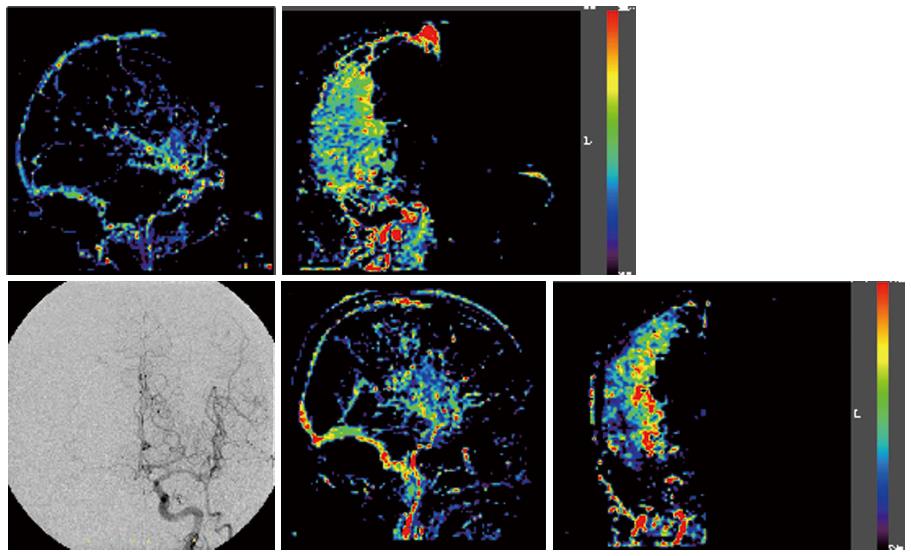
Figure 4 Upper panel: Digital subtraction angiography anterior-posterior view, lateral view showing the blood flow increase ratio, and the anterior-posterior view in Case 3; and Lower panel: Case 4, same series.
Pre-operative digital subtraction angiography in Cases 3 and 4 revealed blood flow to the operative side via the anterior communicating artery. A wide and uniform increase in the area in the middle cerebral artery territory was noted in Case 3. In Case 4, the increase was only noted in a part of the middle cerebral artery perfusion area, especially in the basal ganglia; the rate of increase was high.
- Citation: Wada H, Saito M, Kamada K. Evaluation of changes of intracranial blood flow after carotid artery stenting using digital subtraction angiography flow assessment. World J Radiol 2015; 7(2): 45-51
- URL: https://www.wjgnet.com/1949-8470/full/v7/i2/45.htm
- DOI: https://dx.doi.org/10.4329/wjr.v7.i2.45


