Copyright
©The Author(s) 2015.
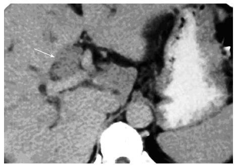
Figure 10 Axial contrast enhanced computed tomography scan in venous phase of papillary type of hilar cholangiocarcinoma showing minimally enhancing intraductal polypoidal lesion (arrow) causing distension of the duct.
- Citation: Madhusudhan KS, Gamanagatti S, Gupta AK. Imaging and interventions in hilar cholangiocarcinoma: A review. World J Radiol 2015; 7(2): 28-44
- URL: https://www.wjgnet.com/1949-8470/full/v7/i2/28.htm
- DOI: https://dx.doi.org/10.4329/wjr.v7.i2.28


