Copyright
©The Author(s) 2015.
World J Radiol. Dec 28, 2015; 7(12): 531-537
Published online Dec 28, 2015. doi: 10.4329/wjr.v7.i12.531
Published online Dec 28, 2015. doi: 10.4329/wjr.v7.i12.531
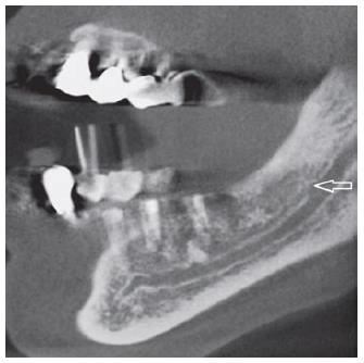
Figure 2 Cone bean computed tomography exam with a white arrow pointing the origin of a forward canal without confluence, bifurcated from the main mandibular canal in the ramus region, and classified as type 3 by Naitoh et al[15] (2009).
- Citation: Castro MAA, Lagravere-Vich MO, Amaral TMP, Abreu MHG, Mesquita RA. Classifications of mandibular canal branching: A review of literature. World J Radiol 2015; 7(12): 531-537
- URL: https://www.wjgnet.com/1949-8470/full/v7/i12/531.htm
- DOI: https://dx.doi.org/10.4329/wjr.v7.i12.531


