Copyright
©The Author(s) 2015.
World J Radiol. Dec 28, 2015; 7(12): 509-520
Published online Dec 28, 2015. doi: 10.4329/wjr.v7.i12.509
Published online Dec 28, 2015. doi: 10.4329/wjr.v7.i12.509
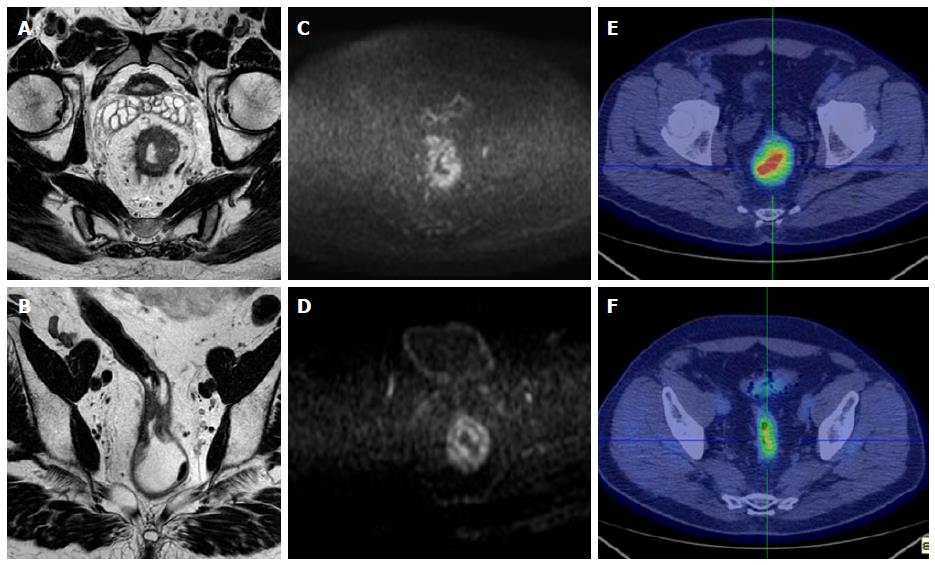
Figure 1 A 72-year-old man with pathologically proven proximal rectal cancer, classified as non-responder after chemoradiation treatment.
A: Pre-CRT T2-weighted axial MR image shows a circumferential pathological rectal mass, with largest thickening from 12 to 6 o’clock position, narrowing rectal lumen and with corresponding mesorectal fat spread around; B: Post-CRT T2- weighted axial MR image shows incomplete decrease in rectal wall thickening, with irregular and inhomogeneous neoplastic tissue still determining lumen narrow; C: Pre-CRT DWI image shows the presence of hyperintense area at the corresponding level of tumor mass (ADC: 0.63 × 10⁻³ mm²/s); D: Post-CRT DWI image demonstrated partial response, with focal hyperintense area still detectable (ADC: 1.25 × 10⁻³ mm²/s); E: Pre-CRT axial pelvic scan examination image demonstrates a significant radiotracer uptake in the hight rectum, corresponding tumor region (SUVmax: 19.4); F: The partial metabolic response is also confirmed by CRT PET/CT scans, in particular a significant tumor uptake is still present (SUVmax: 4.3). CRT: Chemoradiation treatment; DWI: Diffusion weighted imaging; ADC: Apparent diffusion coefficient; SUV: Standardized uptake value.
- Citation: Ippolito D, Fior D, Trattenero C, Ponti ED, Drago S, Guerra L, Franzesi CT, Sironi S. Combined value of apparent diffusion coefficient-standardized uptake value max in evaluation of post-treated locally advanced rectal cancer. World J Radiol 2015; 7(12): 509-520
- URL: https://www.wjgnet.com/1949-8470/full/v7/i12/509.htm
- DOI: https://dx.doi.org/10.4329/wjr.v7.i12.509


