Copyright
©The Author(s) 2015.
World J Radiol. Dec 28, 2015; 7(12): 501-508
Published online Dec 28, 2015. doi: 10.4329/wjr.v7.i12.501
Published online Dec 28, 2015. doi: 10.4329/wjr.v7.i12.501
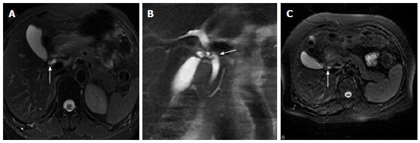
Figure 2 The cystic junction radial orientation.
An FRFSE T2-weighted image (A) shows lateral insertion of the cystic duct (arrow). A coronal SSFSE T2-weighted image (B) shows medial insertion of the cystic duct (arrow). An FRFSE T2-weighted image (C) shows posteroanterior insertion of the cystic duct (arrow). SSFSE: Single-shot fast spin-echo; FRFSE: Fast-recovery fast-spin echo.
- Citation: Peng R, Zhang L, Zhang XM, Chen TW, Yang L, Huang XH, Zhang ZM. Common bile duct diameter in an asymptomatic population: A magnetic resonance imaging study. World J Radiol 2015; 7(12): 501-508
- URL: https://www.wjgnet.com/1949-8470/full/v7/i12/501.htm
- DOI: https://dx.doi.org/10.4329/wjr.v7.i12.501


