Copyright
©The Author(s) 2015.
World J Radiol. Dec 28, 2015; 7(12): 484-493
Published online Dec 28, 2015. doi: 10.4329/wjr.v7.i12.484
Published online Dec 28, 2015. doi: 10.4329/wjr.v7.i12.484
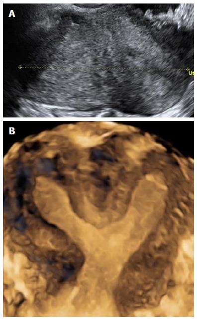
Figure 6 Bicornuate uterus.
A: Transverse 2D ultrasound image of a bicornuate uterus showing the presence of 2 endometrial cavities; B: Coronal 3D ultrasound image of a bicornuate uterus showing external cleft ≥ 1 cm and internal indentation ≥ 1.5 cm. Note the presence of fundal soft tissue separating the 2 uterine cavities, which distinguishes it from uterine didelphys. 3D: Three-dimensional; 2D: Two-dimensional.
- Citation: Wong L, White N, Ramkrishna J, Júnior EA, Meagher S, Costa FDS. Three-dimensional imaging of the uterus: The value of the coronal plane. World J Radiol 2015; 7(12): 484-493
- URL: https://www.wjgnet.com/1949-8470/full/v7/i12/484.htm
- DOI: https://dx.doi.org/10.4329/wjr.v7.i12.484


