Copyright
©The Author(s) 2015.
World J Radiol. Dec 28, 2015; 7(12): 459-474
Published online Dec 28, 2015. doi: 10.4329/wjr.v7.i12.459
Published online Dec 28, 2015. doi: 10.4329/wjr.v7.i12.459
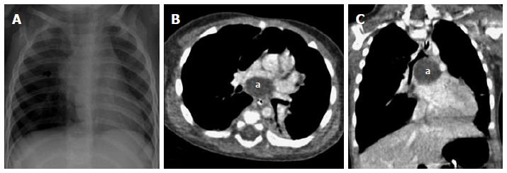
Figure 19 Subcarinal bronchogenic cyst.
Frontal radiograph of chest (A) reveals mediastinal widening (arrow). Contrast enhanced axial (B) and coronal MPR (C) images reveal well defined non-enhancing homogenous fluid attenuation lesion in subcarinal location (a) narrowing the bronchial divisions with resultant subsegmental atelectasis in left lower lobe. MPR: Multiplanar reformation.
- Citation: Jugpal TS, Garg A, Sethi GR, Daga MK, Kumar J. Multi-detector computed tomography imaging of large airway pathology: A pictorial review. World J Radiol 2015; 7(12): 459-474
- URL: https://www.wjgnet.com/1949-8470/full/v7/i12/459.htm
- DOI: https://dx.doi.org/10.4329/wjr.v7.i12.459


