Copyright
©The Author(s) 2015.
World J Radiol. Dec 28, 2015; 7(12): 438-447
Published online Dec 28, 2015. doi: 10.4329/wjr.v7.i12.438
Published online Dec 28, 2015. doi: 10.4329/wjr.v7.i12.438
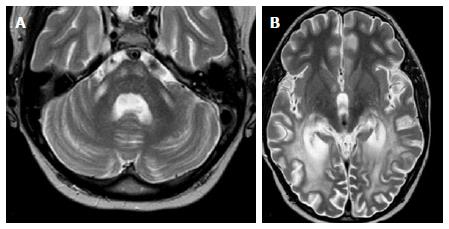
Figure 10 Adrenoleukodystrophy.
A: Axial T2 image shows symmetric increased T2 signal in the MCP and bilateral corticospinal tracts; B: Axial T2 show concomitant symmetric and confluent white matter abnormal signal in bilateral occipito-parietal regions, typical for ADL. Note the preservation of subcortical u-fibers. Images courtesy of Dr. Lily Wang. ADL: Adrenoleukodystrophy; MCP: Middle cerebellar peduncles.
- Citation: Morales H, Tomsick T. Middle cerebellar peduncles: Magnetic resonance imaging and pathophysiologic correlate. World J Radiol 2015; 7(12): 438-447
- URL: https://www.wjgnet.com/1949-8470/full/v7/i12/438.htm
- DOI: https://dx.doi.org/10.4329/wjr.v7.i12.438


