Copyright
©The Author(s) 2015.
World J Radiol. Dec 28, 2015; 7(12): 438-447
Published online Dec 28, 2015. doi: 10.4329/wjr.v7.i12.438
Published online Dec 28, 2015. doi: 10.4329/wjr.v7.i12.438
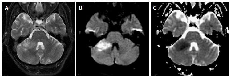
Figure 7 Anterior inferior cerebellar artery infarct.
Patient with acute cerebellar ataxia and right-sided weakness. Axial T2 (A), DWI (B) and ADC maps (C) show well-defined area of high T2 signal and restricted diffusion consistent with ischemia. DWI: Diffusion weighted-imaging.
- Citation: Morales H, Tomsick T. Middle cerebellar peduncles: Magnetic resonance imaging and pathophysiologic correlate. World J Radiol 2015; 7(12): 438-447
- URL: https://www.wjgnet.com/1949-8470/full/v7/i12/438.htm
- DOI: https://dx.doi.org/10.4329/wjr.v7.i12.438


