Copyright
©The Author(s) 2015.
World J Radiol. Nov 28, 2015; 7(11): 394-404
Published online Nov 28, 2015. doi: 10.4329/wjr.v7.i11.394
Published online Nov 28, 2015. doi: 10.4329/wjr.v7.i11.394
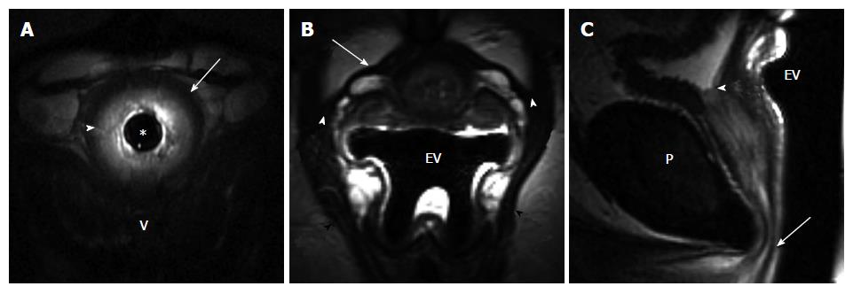
Figure 1 41-year-old woman post one vaginal delivery with episiotomy, body mass index: 46.
7, with occasional stress urinary incontinence. A: Axial T2-weighted image of the mid urethra obtained with 14F endourethral MR coil (TR/TE 4816/68 ms) shows detailed depiction of the urethral sphincter with a hypointense outer layer of striated muscle (arrow) and inner hyperintense smooth muscle layer (arrowhead); B: Axial T2-weighted image at the mid urethra level obtained with endovaginal placement of MRInnervu coil (EV) (TR/TE 3000/92 ms) shows well-defined intact periurethral ligament (arrow) extending between the right and left puborectalis muscle (black arrowheads). Note symmetric, intact ligament attachment (white arrowheads); C: Sagittal T2-weighted image obtained with endovaginal placement of MRInnervu coil (EV) (TR/TE 4000/92 ms) shows normal resting position of the urethra, with distal end of urethral sphincter (arrow) at inferior pubis level (P). Note excellent coaptation of the mucosa at internal meatus/bladder neck level (arrowhead). V: Vagina.
- Citation: Macura KJ, Thompson RE, Bluemke DA, Genadry R. Magnetic resonance imaging in assessment of stress urinary incontinence in women: Parameters differentiating urethral hypermobility and intrinsic sphincter deficiency. World J Radiol 2015; 7(11): 394-404
- URL: https://www.wjgnet.com/1949-8470/full/v7/i11/394.htm
- DOI: https://dx.doi.org/10.4329/wjr.v7.i11.394


