Copyright
©The Author(s) 2015.
World J Radiol. Nov 28, 2015; 7(11): 375-381
Published online Nov 28, 2015. doi: 10.4329/wjr.v7.i11.375
Published online Nov 28, 2015. doi: 10.4329/wjr.v7.i11.375
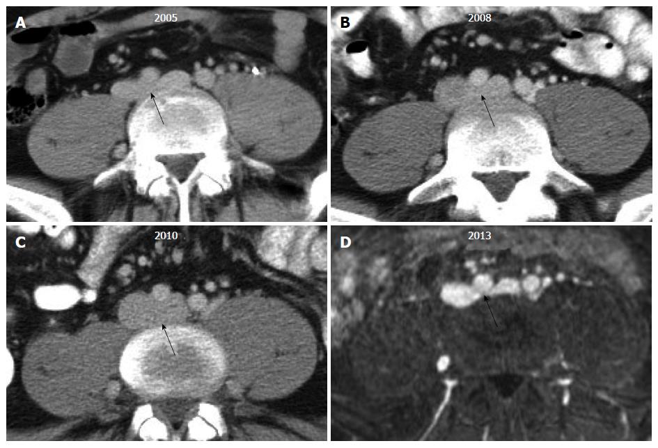
Figure 1 Transverse computed tomography and magnetic resonance images of the proximal left common iliac vein (black arrow) in a single patient across multiple time points illustrate the challenge of diagnosing May-Thurner syndrome.
The degree of venous compression can vary substantially from one imaging study to another based upon the patient’s volume status.
- Citation: Brinegar KN, Sheth RA, Khademhosseini A, Bautista J, Oklu R. Iliac vein compression syndrome: Clinical, imaging and pathologic findings. World J Radiol 2015; 7(11): 375-381
- URL: https://www.wjgnet.com/1949-8470/full/v7/i11/375.htm
- DOI: https://dx.doi.org/10.4329/wjr.v7.i11.375


