Copyright
©The Author(s) 2015.
World J Radiol. Oct 28, 2015; 7(10): 357-360
Published online Oct 28, 2015. doi: 10.4329/wjr.v7.i10.357
Published online Oct 28, 2015. doi: 10.4329/wjr.v7.i10.357
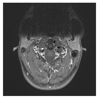
Figure 2 High resolution contrast-enhanced 3T magnetic resonance imaging, fat-saturated gradient echo sequence performed 6 mo following the trauma.
In the contrast-enhanced T1w fat-suppressed magnetic resonance imaging sequence note the contrast enhancement in the periligamentous venous plexus (arrow head). On the left side lower than on the right side. No TS symmetry of the joint space left vs right side (black and white arrow).
- Citation: Kaufmann RA, Marzi I, Vogl TJ. Delayed diagnosis of isolated alar ligament rupture: A case report. World J Radiol 2015; 7(10): 357-360
- URL: https://www.wjgnet.com/1949-8470/full/v7/i10/357.htm
- DOI: https://dx.doi.org/10.4329/wjr.v7.i10.357


