Copyright
©The Author(s) 2015.
World J Radiol. Oct 28, 2015; 7(10): 329-335
Published online Oct 28, 2015. doi: 10.4329/wjr.v7.i10.329
Published online Oct 28, 2015. doi: 10.4329/wjr.v7.i10.329
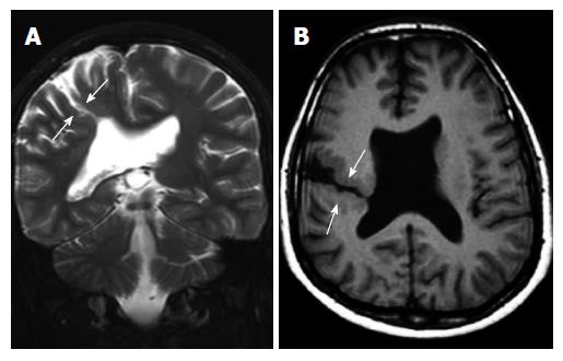
Figure 11 A 12-year-old boy with schizencephaly.
T2-weighted coronal (A) and T1-weighted axial (B) images show oblique gray matter lined holohemispheric cleft (arrows) extending into the lateral ventricle that suggest open lip type schizencephaly with agenesis of septum pellucidum.
- Citation: Battal B, Ince S, Akgun V, Kocaoglu M, Ozcan E, Tasar M. Malformations of cortical development: 3T magnetic resonance imaging features. World J Radiol 2015; 7(10): 329-335
- URL: https://www.wjgnet.com/1949-8470/full/v7/i10/329.htm
- DOI: https://dx.doi.org/10.4329/wjr.v7.i10.329


