Copyright
©The Author(s) 2015.
World J Radiol. Oct 28, 2015; 7(10): 329-335
Published online Oct 28, 2015. doi: 10.4329/wjr.v7.i10.329
Published online Oct 28, 2015. doi: 10.4329/wjr.v7.i10.329
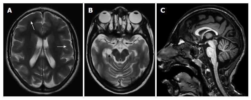
Figure 3 An 8-year-old girl with microlissencephaly.
Axial T2-weighted images (A, B) show reduction in the number and depth of the sulcus, thickened cortex (white arrows) with choroidal fissure cyst (black arrow). Thinning of posterior parts of the corpus callosum is also seen on sagittal three-dimensional (3D) T1-weighted image (C).
- Citation: Battal B, Ince S, Akgun V, Kocaoglu M, Ozcan E, Tasar M. Malformations of cortical development: 3T magnetic resonance imaging features. World J Radiol 2015; 7(10): 329-335
- URL: https://www.wjgnet.com/1949-8470/full/v7/i10/329.htm
- DOI: https://dx.doi.org/10.4329/wjr.v7.i10.329


