Copyright
©The Author(s) 2015.
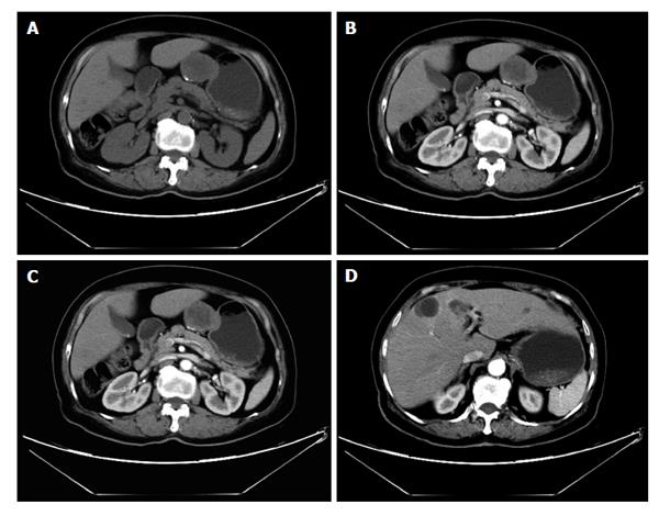
Figure 11 A 76-year-old female with gastrointestinal stromal tumors.
Axial plain (A) shows an round, exophytic soft tissue mass, appearing as a dominant mass extrinsic to the wall of the stomach, contrast-enhanced computed tomography arterial (B) and venous phase (C) scan shows slightly enhancing, occasionally with coarse calcification. Liver metastases were shown (D).
- Citation: Li YZ, Wu PH. Conventional radiological strategy of common gastrointestinal neoplasms. World J Radiol 2015; 7(1): 7-16
- URL: https://www.wjgnet.com/1949-8470/full/v7/i1/7.htm
- DOI: https://dx.doi.org/10.4329/wjr.v7.i1.7


