Copyright
©The Author(s) 2015.
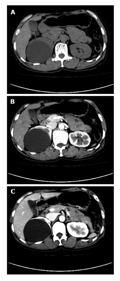
Figure 10 A 60-year-old female patient with mucosa-associated lymphoid tissue in gastric antrum.
The tumor reveals as segmental, smooth homogeneous wall thickening on non-contrast computed tomography (A), minimal enhancement on arterial (B) and venous (C) phase enhancement.
- Citation: Li YZ, Wu PH. Conventional radiological strategy of common gastrointestinal neoplasms. World J Radiol 2015; 7(1): 7-16
- URL: https://www.wjgnet.com/1949-8470/full/v7/i1/7.htm
- DOI: https://dx.doi.org/10.4329/wjr.v7.i1.7


