Copyright
©The Author(s) 2015.
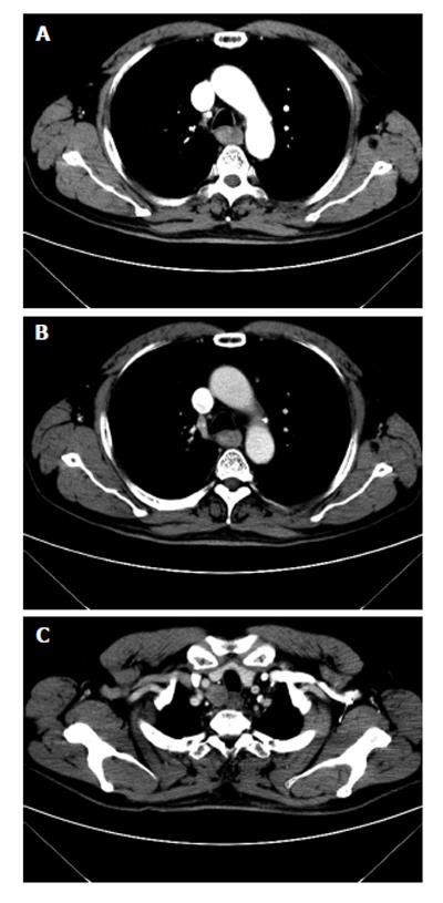
Figure 4 A 61-year-old male with squamous cell carcinoma in the first third section of esophagus.
At computed tomography, esophageal cancer shows a soft tissue mass, with irregular luminal narrowing. The located wall thickening was asymmetric. The lesion shows moderate arterial phase enhancement (A) and slight venous phase enhancement (B). The enlarged lymph node metastasis is shown in the upper right paratracheal (C).
- Citation: Li YZ, Wu PH. Conventional radiological strategy of common gastrointestinal neoplasms. World J Radiol 2015; 7(1): 7-16
- URL: https://www.wjgnet.com/1949-8470/full/v7/i1/7.htm
- DOI: https://dx.doi.org/10.4329/wjr.v7.i1.7


