Copyright
©The Author(s) 2015.
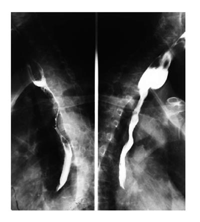
Figure 2 A 60-year-old male with squamous cell carcinoma.
On barium esophagography, infiltration in the upper third of esophageal carcinoma, with irregular luminal narrowing, mucosal destruction, dilatation and abrupt proximal borders. Prestenotic dilatation is also present.
- Citation: Li YZ, Wu PH. Conventional radiological strategy of common gastrointestinal neoplasms. World J Radiol 2015; 7(1): 7-16
- URL: https://www.wjgnet.com/1949-8470/full/v7/i1/7.htm
- DOI: https://dx.doi.org/10.4329/wjr.v7.i1.7


