Copyright
©The Author(s) 2015.
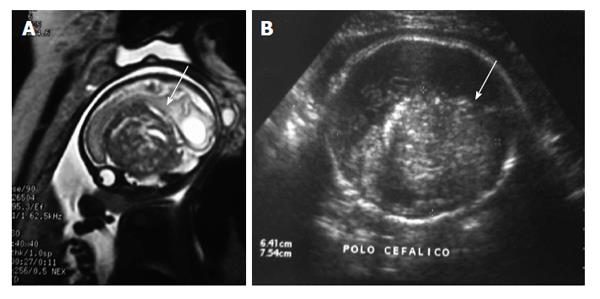
Figure 4 Craniopharyngioma.
(A) Fetal magnetic resonance imaging (T2 haste, sagittal plane) and (B) ultrasound (axial plane) at 28 wk of gestation showing a heterogeneous and hyperechogenic suprasellar lesion and hydrocephalus (white arrows). Histology confirmed craniopharyngioma.
- Citation: Milani HJ, Araujo Júnior E, Cavalheiro S, Oliveira PS, Hisaba WJ, Barreto EQS, Barbosa MM, Nardozza LM, Moron AF. Fetal brain tumors: Prenatal diagnosis by ultrasound and magnetic resonance imaging. World J Radiol 2015; 7(1): 17-21
- URL: https://www.wjgnet.com/1949-8470/full/v7/i1/17.htm
- DOI: https://dx.doi.org/10.4329/wjr.v7.i1.17


