Copyright
©The Author(s) 2015.
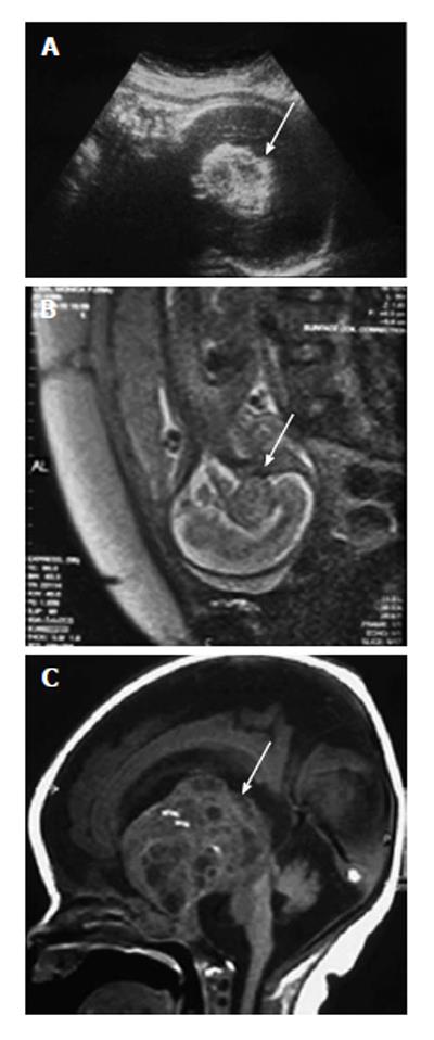
Figure 3 Teratoma.
A: Ultrasound (US) at 29 wk of gestation (median sagittal plane) showing heterogeneous and hyperechogenic lesion in the suprasellar region (white arrow); B: Fetal magnetic resonance imaging (T2 haste, sagittal plane) at 29 wk of gestation confirming aspects observed on US (white arrow); C: Magnetic resonance imaging (T1 Fast Spin Echo, sagittal plane) 3 d after birth showing suprasellar lesion with solid and cystic components distorting brain anatomy (white arrow). Histology confirmed teratoma.
- Citation: Milani HJ, Araujo Júnior E, Cavalheiro S, Oliveira PS, Hisaba WJ, Barreto EQS, Barbosa MM, Nardozza LM, Moron AF. Fetal brain tumors: Prenatal diagnosis by ultrasound and magnetic resonance imaging. World J Radiol 2015; 7(1): 17-21
- URL: https://www.wjgnet.com/1949-8470/full/v7/i1/17.htm
- DOI: https://dx.doi.org/10.4329/wjr.v7.i1.17


