Copyright
©2014 Baishideng Publishing Group Inc.
World J Radiol. Sep 28, 2014; 6(9): 737-740
Published online Sep 28, 2014. doi: 10.4329/wjr.v6.i9.737
Published online Sep 28, 2014. doi: 10.4329/wjr.v6.i9.737
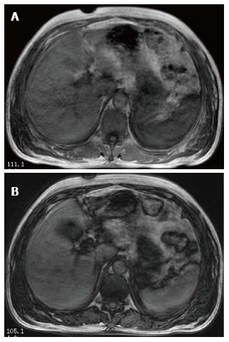
Figure 3 Axial LAVA-Flex T1 Weighted In-Phase image (A) and gradient echo T1 Weighted Out-of-Phase image (B) of the abdomen show decreased signal intensity of the spleen on In-Phase image (A) as compared to Out-of-Phase image (B).
- Citation: Das SK, Zeng LC, Li B, Niu XK, Wang JL, Bhetuwal A, Yang HF. Magnetic resonance imaging correlates of bee sting induced multiple organ dysfunction syndrome: A case report. World J Radiol 2014; 6(9): 737-740
- URL: https://www.wjgnet.com/1949-8470/full/v6/i9/737.htm
- DOI: https://dx.doi.org/10.4329/wjr.v6.i9.737


