Copyright
©2014 Baishideng Publishing Group Inc.
World J Radiol. Sep 28, 2014; 6(9): 730-736
Published online Sep 28, 2014. doi: 10.4329/wjr.v6.i9.730
Published online Sep 28, 2014. doi: 10.4329/wjr.v6.i9.730
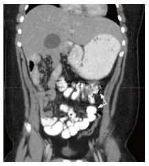
Figure 4 This coronal view of an abdominal computed tomography illustrates the terminal ileum and cecum (white arrows).
Positioning of the cecum in the left hemi-abdomen is suggestive of malrotation.
- Citation: Tackett JJ, Muise ED, Cowles RA. Malrotation: Current strategies navigating the radiologic diagnosis of a surgical emergency. World J Radiol 2014; 6(9): 730-736
- URL: https://www.wjgnet.com/1949-8470/full/v6/i9/730.htm
- DOI: https://dx.doi.org/10.4329/wjr.v6.i9.730


