Copyright
©2014 Baishideng Publishing Group Inc.
World J Radiol. Sep 28, 2014; 6(9): 657-668
Published online Sep 28, 2014. doi: 10.4329/wjr.v6.i9.657
Published online Sep 28, 2014. doi: 10.4329/wjr.v6.i9.657
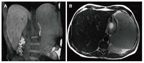
Figure 1 Hepatosplenomegaly.
Coronal T1 WI (A) and axial T2 WI (B) images in a male type 1 GD patient with N370S/N370S genotype demonstrate marked hepatosplenomegaly. The liver volume measured 3235 cc. The spleen volume measured 2923 cc.
- Citation: Simpson WL, Hermann G, Balwani M. Imaging of Gaucher disease. World J Radiol 2014; 6(9): 657-668
- URL: https://www.wjgnet.com/1949-8470/full/v6/i9/657.htm
- DOI: https://dx.doi.org/10.4329/wjr.v6.i9.657


