Copyright
©2014 Baishideng Publishing Group Inc.
World J Radiol. Sep 28, 2014; 6(9): 636-642
Published online Sep 28, 2014. doi: 10.4329/wjr.v6.i9.636
Published online Sep 28, 2014. doi: 10.4329/wjr.v6.i9.636
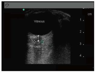
Figure 8 Optic nerve sheath diameter.
Using a 7.5-MHz linear probe on the closed upper eyelid, the optic nerve was visualized as a linear hypoechoic structure with clearly defined margins posterior to the globe. Sheath diameter was measured 3 mm behind the globe. ONSD: Optic nerve sheath diameter.
- Citation: Caricato A, Pitoni S, Montini L, Bocci MG, Annetta P, Antonelli M. Echography in brain imaging in intensive care unit: State of the art. World J Radiol 2014; 6(9): 636-642
- URL: https://www.wjgnet.com/1949-8470/full/v6/i9/636.htm
- DOI: https://dx.doi.org/10.4329/wjr.v6.i9.636


