Copyright
©2014 Baishideng Publishing Group Inc.
World J Radiol. Sep 28, 2014; 6(9): 636-642
Published online Sep 28, 2014. doi: 10.4329/wjr.v6.i9.636
Published online Sep 28, 2014. doi: 10.4329/wjr.v6.i9.636
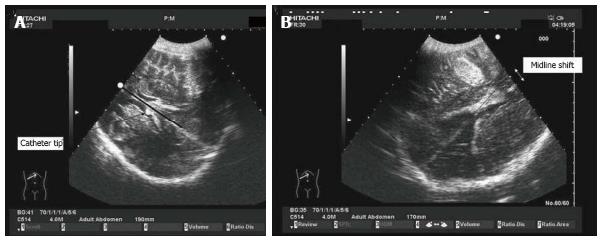
Figure 7 Midline shift in decompressive craniectomy.
Images obtained through decompressive craniectomy. Midline was identified with the interventricular line. The distance between the extension of falx and the interventricular line was measured as midline shift (MLS). In the case on the left (A), the extension of the falx exactly overlaps with the interventricular line (dark line). On the right (B), MLS caused by a temporal hematoma is shown.
- Citation: Caricato A, Pitoni S, Montini L, Bocci MG, Annetta P, Antonelli M. Echography in brain imaging in intensive care unit: State of the art. World J Radiol 2014; 6(9): 636-642
- URL: https://www.wjgnet.com/1949-8470/full/v6/i9/636.htm
- DOI: https://dx.doi.org/10.4329/wjr.v6.i9.636


