Copyright
©2014 Baishideng Publishing Group Inc.
World J Radiol. Sep 28, 2014; 6(9): 636-642
Published online Sep 28, 2014. doi: 10.4329/wjr.v6.i9.636
Published online Sep 28, 2014. doi: 10.4329/wjr.v6.i9.636
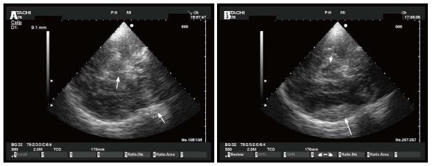
Figure 5 Epidural hematoma in echography.
A: Epidural hematoma. A small epidural hematoma (arrow on the right) is shown as an hyperechogenic image just inside the skull. Arrow on the left indicates mesencephalon; B: Epidural hematoma. The same epidural hematoma of Figure A (arrow) 1 h later. White arrow indicates mesencephalon.
- Citation: Caricato A, Pitoni S, Montini L, Bocci MG, Annetta P, Antonelli M. Echography in brain imaging in intensive care unit: State of the art. World J Radiol 2014; 6(9): 636-642
- URL: https://www.wjgnet.com/1949-8470/full/v6/i9/636.htm
- DOI: https://dx.doi.org/10.4329/wjr.v6.i9.636


