Copyright
©2014 Baishideng Publishing Group Inc.
World J Radiol. Aug 28, 2014; 6(8): 613-618
Published online Aug 28, 2014. doi: 10.4329/wjr.v6.i8.613
Published online Aug 28, 2014. doi: 10.4329/wjr.v6.i8.613
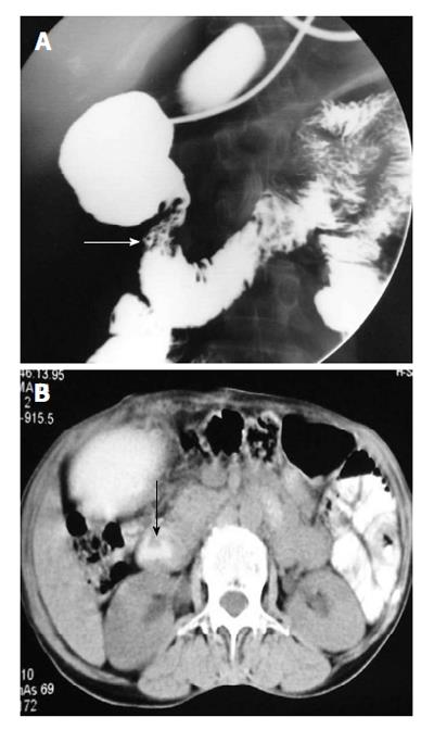
Figure 5 Upper gastrointestinal barium study (A) demonstrates smooth circumferential extrinsic narrowing of the second part of the duodenum (arrow), axial computed tomography image (B) shows pancreatic parenchyma incompletely surrounding the duodenum (arrow), features suggestive of partial annular pancreas.
- Citation: Gupta P, Debi U, Sinha SK, Prasad KK. Upper gastrointestinal barium evaluation of duodenal pathology: A pictorial review. World J Radiol 2014; 6(8): 613-618
- URL: https://www.wjgnet.com/1949-8470/full/v6/i8/613.htm
- DOI: https://dx.doi.org/10.4329/wjr.v6.i8.613


