Copyright
©2014 Baishideng Publishing Group Inc.
World J Radiol. Aug 28, 2014; 6(8): 583-588
Published online Aug 28, 2014. doi: 10.4329/wjr.v6.i8.583
Published online Aug 28, 2014. doi: 10.4329/wjr.v6.i8.583
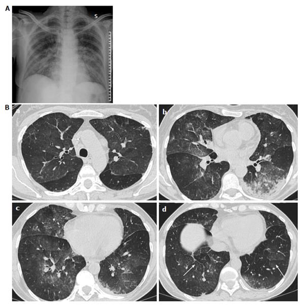
Figure 2 Photograph.
A: Chest X-ray shows subtile patchy ground glass opacities of the middle inferior lung fields; B: Computed tomography scans of the same patient as Figure 4 shows patchy ground glass opacities (a, b and c) with interlobar septal thickening (arrows in d). References: Institute of Radiology, Department of Clinical and Biological Sciences of University of Turin, AOU S.Luigi Gonzaga, Regione Gonzole 10, 10043 Orbassano, Torino, Italy.
- Citation: Cardinale L, Asteggiano F, Moretti F, Torre F, Ulisciani S, Fava C, Rege-Cambrin G. Pathophysiology, clinical features and radiological findings of differentiation syndrome/all-trans-retinoic acid syndrome. World J Radiol 2014; 6(8): 583-588
- URL: https://www.wjgnet.com/1949-8470/full/v6/i8/583.htm
- DOI: https://dx.doi.org/10.4329/wjr.v6.i8.583


