Copyright
©2014 Baishideng Publishing Group Inc.
World J Radiol. Aug 28, 2014; 6(8): 567-582
Published online Aug 28, 2014. doi: 10.4329/wjr.v6.i8.567
Published online Aug 28, 2014. doi: 10.4329/wjr.v6.i8.567
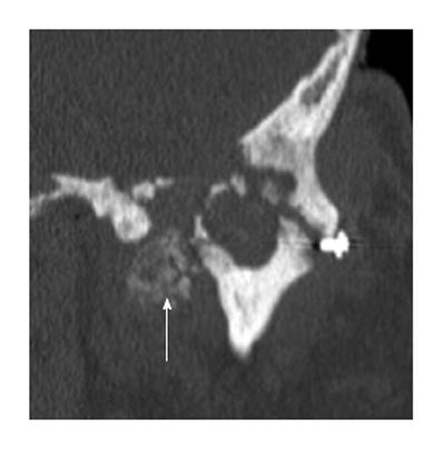
Figure 18 Calcium pyrophosphate dehydrate deposition disease.
Coronal reformation of the axial dataset demonstrates destruction of the left temporomandibular joint with erosion and deformity of both the mandibular condyle and the glenoid fossa. There is extensive extensive calcium pyrophosphate dehydrate deposition disease medial to the joint space (arrow).
- Citation: Bag AK, Gaddikeri S, Singhal A, Hardin S, Tran BD, Medina JA, Curé JK. Imaging of the temporomandibular joint: An update. World J Radiol 2014; 6(8): 567-582
- URL: https://www.wjgnet.com/1949-8470/full/v6/i8/567.htm
- DOI: https://dx.doi.org/10.4329/wjr.v6.i8.567


