Copyright
©2014 Baishideng Publishing Group Inc.
World J Radiol. Aug 28, 2014; 6(8): 567-582
Published online Aug 28, 2014. doi: 10.4329/wjr.v6.i8.567
Published online Aug 28, 2014. doi: 10.4329/wjr.v6.i8.567
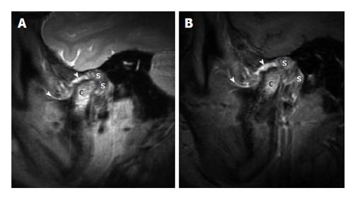
Figure 16 Juvenile idiopathic arthritis.
A: Sagittal proton density weighted magnetic resonance imaging (MRI) in the closed mouth position demonstrates increased signal at the mandibular condyle (the letter, c), extensive thickening of the synovium (the letter, s) in the retrodiscal regions. It can be noted that the thickening and increased signal of the synovium at other places (arrowheads); B: Sagittal fat suppressed post contrast T1 weighted MRI in the closed mouth position demonstrates enhancement of signal at the mandibular condyle (the letter, c), enhancement and extensive thickening of the synovium (the letter, s) in the retrodiscal regions. There is thickening and enhancement of the synovium at other places (arrowheads).
- Citation: Bag AK, Gaddikeri S, Singhal A, Hardin S, Tran BD, Medina JA, Curé JK. Imaging of the temporomandibular joint: An update. World J Radiol 2014; 6(8): 567-582
- URL: https://www.wjgnet.com/1949-8470/full/v6/i8/567.htm
- DOI: https://dx.doi.org/10.4329/wjr.v6.i8.567


