Copyright
©2014 Baishideng Publishing Group Inc.
World J Radiol. Aug 28, 2014; 6(8): 567-582
Published online Aug 28, 2014. doi: 10.4329/wjr.v6.i8.567
Published online Aug 28, 2014. doi: 10.4329/wjr.v6.i8.567
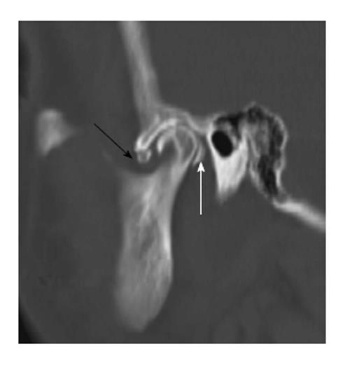
Figure 14 Loose bodies.
Sagittal reformation of the axial dataset demonstrates multiple “loose bodies” in the joint cavities, anteroinferior to the articular eminence (black arrow) and immediately posterior to the mandibular condyle (white arrow).
- Citation: Bag AK, Gaddikeri S, Singhal A, Hardin S, Tran BD, Medina JA, Curé JK. Imaging of the temporomandibular joint: An update. World J Radiol 2014; 6(8): 567-582
- URL: https://www.wjgnet.com/1949-8470/full/v6/i8/567.htm
- DOI: https://dx.doi.org/10.4329/wjr.v6.i8.567


