Copyright
©2014 Baishideng Publishing Group Inc.
World J Radiol. Aug 28, 2014; 6(8): 567-582
Published online Aug 28, 2014. doi: 10.4329/wjr.v6.i8.567
Published online Aug 28, 2014. doi: 10.4329/wjr.v6.i8.567
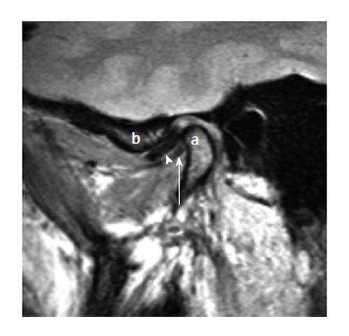
Figure 12 Double disk sign (thickening of the lateral pterygoid muscle).
Sagittal closed mouth proton density image demonstrates anterior displacement of the disk (arrow head). The thickened lateral pterygoid muscle near the mandibular condylar (the letter, a) attachment appear as linear hypointense structure (white arrow) inferior to the disk in the same orientation giving the appearance of “double disk”. The articular eminence is denoted with letter “b”.
- Citation: Bag AK, Gaddikeri S, Singhal A, Hardin S, Tran BD, Medina JA, Curé JK. Imaging of the temporomandibular joint: An update. World J Radiol 2014; 6(8): 567-582
- URL: https://www.wjgnet.com/1949-8470/full/v6/i8/567.htm
- DOI: https://dx.doi.org/10.4329/wjr.v6.i8.567


