Copyright
©2014 Baishideng Publishing Group Inc.
World J Radiol. Aug 28, 2014; 6(8): 567-582
Published online Aug 28, 2014. doi: 10.4329/wjr.v6.i8.567
Published online Aug 28, 2014. doi: 10.4329/wjr.v6.i8.567
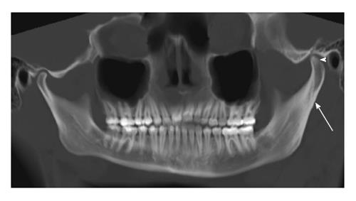
Figure 6 Condylar hyperplasia.
Panoramic reformation of the source computed tomography data including both the temporomandibular joints of a young patient demonstrates hyperplasia of the left condyle (arrowhead) in comparison to the right side. Associated hypertrophy of the ramus and the neck (arrow) of the left hemi-mandible is also noted.
- Citation: Bag AK, Gaddikeri S, Singhal A, Hardin S, Tran BD, Medina JA, Curé JK. Imaging of the temporomandibular joint: An update. World J Radiol 2014; 6(8): 567-582
- URL: https://www.wjgnet.com/1949-8470/full/v6/i8/567.htm
- DOI: https://dx.doi.org/10.4329/wjr.v6.i8.567


