Copyright
©2014 Baishideng Publishing Group Inc.
World J Radiol. Jul 28, 2014; 6(7): 493-501
Published online Jul 28, 2014. doi: 10.4329/wjr.v6.i7.493
Published online Jul 28, 2014. doi: 10.4329/wjr.v6.i7.493
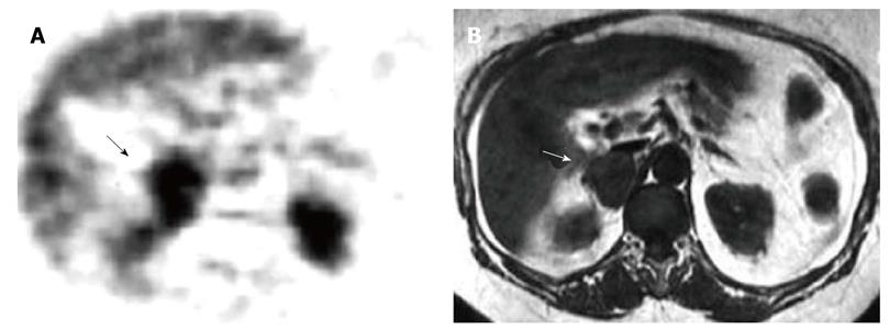
Figure 3 Right adrenal metastasis by melanoma.
A: Axial fluorine-18 fludeoxyglucose-positron emission tomography scan detects abnormal focal uptake in the right adrenal region (black arrow) where a large adrenal metastasis was detected by magnetic resonance (MR); diffuse normal liver tracer activity is detectable; physiologic tracer activity is also detectable in kidneys; B: T1-weighted axial MR detects a right adrenal tumor hypointense compared to liver signal intensity (white arrow).
- Citation: Maurea S, Mainenti PP, Romeo V, Mollica C, Salvatore M. Nuclear imaging to characterize adrenal tumors: Comparison with MRI. World J Radiol 2014; 6(7): 493-501
- URL: https://www.wjgnet.com/1949-8470/full/v6/i7/493.htm
- DOI: https://dx.doi.org/10.4329/wjr.v6.i7.493


