Copyright
©2014 Baishideng Publishing Group Inc.
World J Radiol. Jul 28, 2014; 6(7): 399-408
Published online Jul 28, 2014. doi: 10.4329/wjr.v6.i7.399
Published online Jul 28, 2014. doi: 10.4329/wjr.v6.i7.399
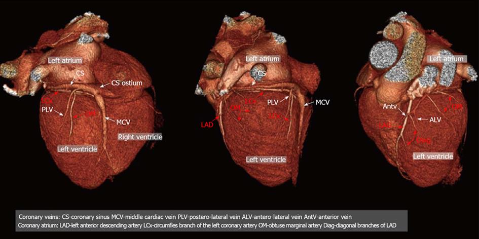
Figure 1 Example of the three-dimensional (3D) anatomy of coronary vessels (arteries and veins).
Posterior, lateral antero-lateral view of the heart; 3D volume rendering projections. CS: Coronary sinus; MCV: Middle cardiac vein; PLV: Postero-lateral vein; ALV: Antero-lateral vein; AntV: Anterior vein; LAD: Left anterior descending artery; LCx: Circumfles branch of the left coronary artery; OM: Obtuse marginal artery; Diag: Diagonal branches of LAD.
- Citation: Mlynarski R, Mlynarska A, Sosnowski M. Coronary venous system in cardiac computer tomography: Visualization, classification and role. World J Radiol 2014; 6(7): 399-408
- URL: https://www.wjgnet.com/1949-8470/full/v6/i7/399.htm
- DOI: https://dx.doi.org/10.4329/wjr.v6.i7.399


