Copyright
©2014 Baishideng Publishing Group Inc.
World J Radiol. Jun 28, 2014; 6(6): 355-365
Published online Jun 28, 2014. doi: 10.4329/wjr.v6.i6.355
Published online Jun 28, 2014. doi: 10.4329/wjr.v6.i6.355
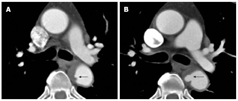
Figure 9 Computed tomography aortogram in a 75-year-old man shows an intramural hematoma (note the eccentric wall thickening in the descending aorta) with intimal surface defect (A: arrow).
At one year follow up it has progressed into a frank penetrating ulcer (B: arrow).
- Citation: Hallinan JTPD, Anil G. Multi-detector computed tomography in the diagnosis and management of acute aortic syndromes. World J Radiol 2014; 6(6): 355-365
- URL: https://www.wjgnet.com/1949-8470/full/v6/i6/355.htm
- DOI: https://dx.doi.org/10.4329/wjr.v6.i6.355


