Copyright
©2014 Baishideng Publishing Group Inc.
World J Radiol. Jun 28, 2014; 6(6): 355-365
Published online Jun 28, 2014. doi: 10.4329/wjr.v6.i6.355
Published online Jun 28, 2014. doi: 10.4329/wjr.v6.i6.355
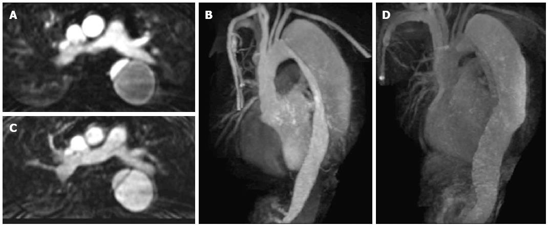
Figure 3 Axial contrast enhanced magnetic resonance aortogram of the same patient as in Figure 2 with type B aortic dissection.
In the arterial phase image (A: Axial; B: Coronal 3D reconstruction; 30 s post injection) the true lumen is of small calibre and shows early intense contrast enhancement compared to the larger false lumen. In the second delayed phase (70 s post injection) the enhancement between the lumens becomes more similar (C: Axial; D: Coronal 3D reconstruction).
- Citation: Hallinan JTPD, Anil G. Multi-detector computed tomography in the diagnosis and management of acute aortic syndromes. World J Radiol 2014; 6(6): 355-365
- URL: https://www.wjgnet.com/1949-8470/full/v6/i6/355.htm
- DOI: https://dx.doi.org/10.4329/wjr.v6.i6.355


