Copyright
©2014 Baishideng Publishing Group Inc.
World J Radiol. Jun 28, 2014; 6(6): 344-354
Published online Jun 28, 2014. doi: 10.4329/wjr.v6.i6.344
Published online Jun 28, 2014. doi: 10.4329/wjr.v6.i6.344
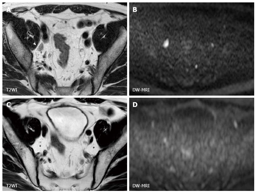
Figure 2 Magnetic resonance images of a 45-year-old man with muscle-invasive bladder cancer (urothelial cancer, stage cT3N1) before and after chemoradiotherapy.
A: Before CRT, an enlarged right external iliac lymph node (arrow head) is visible on T2WI; B: The lymph node on the corresponding DW-MRI shows a hyperintense signal; C and D: After CRT, size reduction on T2WI (C) and signal attenuation on DW-MRI (D) in lymph node is evident, consistent with the expected treatment response. CRT: Chemoradiotherapy; DW-MRI: Diffusion-weighted magnetic resonance imaging.
- Citation: Yoshida S, Koga F, Kobayashi S, Tanaka H, Satoh S, Fujii Y, Kihara K. Diffusion-weighted magnetic resonance imaging in management of bladder cancer, particularly with multimodal bladder-sparing strategy. World J Radiol 2014; 6(6): 344-354
- URL: https://www.wjgnet.com/1949-8470/full/v6/i6/344.htm
- DOI: https://dx.doi.org/10.4329/wjr.v6.i6.344


