Copyright
©2014 Baishideng Publishing Group Inc.
World J Radiol. Jun 28, 2014; 6(6): 344-354
Published online Jun 28, 2014. doi: 10.4329/wjr.v6.i6.344
Published online Jun 28, 2014. doi: 10.4329/wjr.v6.i6.344
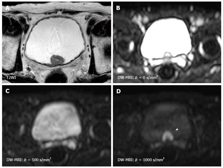
Figure 1 Magnetic resonance images of a 79-year-old man with non-muscle invasive bladder cancer (urothelial cancer, stage pTa, grade 2 > 3).
A: T2WI shows a hypointense tumor at the trigone; B: The signal intensity of diffusion-weighted magnetic resonance imaging depends on both water diffusion and the T2 relaxation time; C: Due to the very long T2 relaxation time of urine, the signal of the urine in the bladder remains high on the diffusion-weighted magnetic resonance imaging with a b-value of 500 s/mm2. This is known as the “T2 shine-through effect”; D: Using a b-value of 1000 s/mm2 decreases the signal of the urine, as well as those of the seminal vesicles, while the bladder cancer (arrow head) shows little signal attenuation with the increased b-value.
- Citation: Yoshida S, Koga F, Kobayashi S, Tanaka H, Satoh S, Fujii Y, Kihara K. Diffusion-weighted magnetic resonance imaging in management of bladder cancer, particularly with multimodal bladder-sparing strategy. World J Radiol 2014; 6(6): 344-354
- URL: https://www.wjgnet.com/1949-8470/full/v6/i6/344.htm
- DOI: https://dx.doi.org/10.4329/wjr.v6.i6.344


