Copyright
©2014 Baishideng Publishing Group Inc.
World J Radiol. Jun 28, 2014; 6(6): 313-328
Published online Jun 28, 2014. doi: 10.4329/wjr.v6.i6.313
Published online Jun 28, 2014. doi: 10.4329/wjr.v6.i6.313
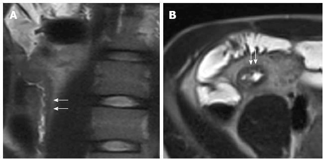
Figure 14 Coronal (A), transverse (B) T2-weighted images show thickened, inflamed segments of the terminal ileum with deep ulcers seen as high-contrast protrusions within bowel wall (arrows).
- Citation: Casciani E, Vincentiis CD, Polettini E, Masselli G, Nardo GD, Civitelli F, Cucchiara S, Gualdi GF. Imaging of the small bowel: Crohn’s disease in paediatric patients. World J Radiol 2014; 6(6): 313-328
- URL: https://www.wjgnet.com/1949-8470/full/v6/i6/313.htm
- DOI: https://dx.doi.org/10.4329/wjr.v6.i6.313


