Copyright
©2014 Baishideng Publishing Group Inc.
World J Radiol. Jun 28, 2014; 6(6): 284-300
Published online Jun 28, 2014. doi: 10.4329/wjr.v6.i6.284
Published online Jun 28, 2014. doi: 10.4329/wjr.v6.i6.284
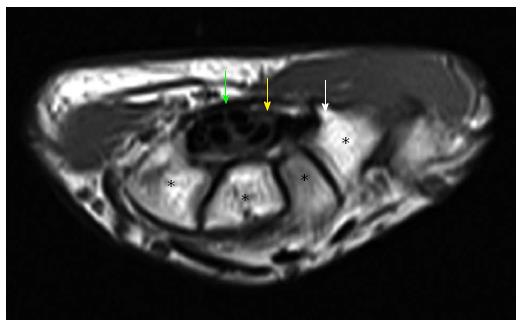
Figure 8 Axial T1W image of carpal tunnel at the level of tunnel outlet shows bony part of carpal tunnel as intermediate signal intensity composed from left to right hamate, capitates, trapezoid, trapezium.
White arrow shows hook of hamate, yellow arrow shows median nerve, green arrow shows flexor retinaculum. Asterisks indicate carpal bones.
- Citation: Ghasemi-rad M, Nosair E, Vegh A, Mohammadi A, Akkad A, Lesha E, Mohammadi MH, Sayed D, Davarian A, Maleki-Miyandoab T, Hasan A. A handy review of carpal tunnel syndrome: From anatomy to diagnosis and treatment. World J Radiol 2014; 6(6): 284-300
- URL: https://www.wjgnet.com/1949-8470/full/v6/i6/284.htm
- DOI: https://dx.doi.org/10.4329/wjr.v6.i6.284


