Copyright
©2014 Baishideng Publishing Group Inc.
World J Radiol. Jun 28, 2014; 6(6): 284-300
Published online Jun 28, 2014. doi: 10.4329/wjr.v6.i6.284
Published online Jun 28, 2014. doi: 10.4329/wjr.v6.i6.284
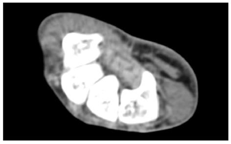
Figure 3 Axial computed tomography scan shows bony part of carpal tunnel at the level of outlet.
Bony structures from left to right are HAMATE, CAPITATE, TRAPEZOID, TRAPEZIUM. FR (arrow) b and flexor tendons can be detected by computed tomography scan.
- Citation: Ghasemi-rad M, Nosair E, Vegh A, Mohammadi A, Akkad A, Lesha E, Mohammadi MH, Sayed D, Davarian A, Maleki-Miyandoab T, Hasan A. A handy review of carpal tunnel syndrome: From anatomy to diagnosis and treatment. World J Radiol 2014; 6(6): 284-300
- URL: https://www.wjgnet.com/1949-8470/full/v6/i6/284.htm
- DOI: https://dx.doi.org/10.4329/wjr.v6.i6.284


