Copyright
©2014 Baishideng Publishing Group Inc.
World J Radiol. Jun 28, 2014; 6(6): 274-283
Published online Jun 28, 2014. doi: 10.4329/wjr.v6.i6.274
Published online Jun 28, 2014. doi: 10.4329/wjr.v6.i6.274
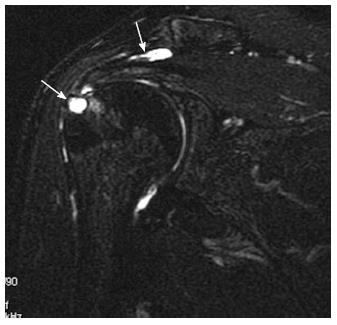
Figure 16 Bony degenerative changes.
Coronal oblique fat-sat T2-weighted image showing focal fluid-like high signal within the distal supraspinatus tendon fibres reported as partial capsular surface tear with subcortical cystic erosion of the greater tuberosity at the rotor cuff insertion (thin arrow). Also note fluid signal within the subacromial subdeltoid bursa reported as bursitis (thick arrow).
- Citation: Tawfik AM, El-Morsy A, Badran MA. Rotator cuff disorders: How to write a surgically relevant magnetic resonance imaging report? World J Radiol 2014; 6(6): 274-283
- URL: https://www.wjgnet.com/1949-8470/full/v6/i6/274.htm
- DOI: https://dx.doi.org/10.4329/wjr.v6.i6.274


