Copyright
©2014 Baishideng Publishing Group Inc.
World J Radiol. Jun 28, 2014; 6(6): 238-251
Published online Jun 28, 2014. doi: 10.4329/wjr.v6.i6.238
Published online Jun 28, 2014. doi: 10.4329/wjr.v6.i6.238
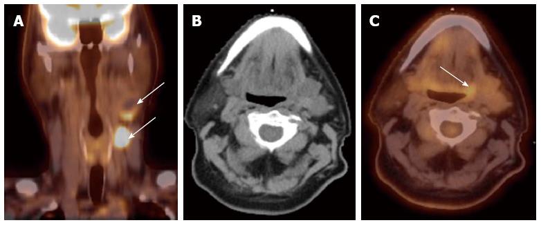
Figure 1 Computed tomography.
A: A patient presented with multiple left neck nodes (arrows); B: The conventional methods as well as the computed tomography (CT) imaging could not identify the primary tumor; C: A positron emission tomography/CT scan showed increased fluorodeoxyglucose uptake in the left base of tongue (arrow) and a directed biopsy of this area confirmed the primary site.
- Citation: Siddiqui F, Yao M. Application of fluorodeoxyglucose positron emission tomography in the management of head and neck cancers. World J Radiol 2014; 6(6): 238-251
- URL: https://www.wjgnet.com/1949-8470/full/v6/i6/238.htm
- DOI: https://dx.doi.org/10.4329/wjr.v6.i6.238


